
Airway conditions and thoracic biopsies
Diagnostic assessment of the trachea, bronchi and tissues within the chest.
Draft awaiting medical review · General information; not a substitute for an individual consultation and assessment.
Content updated: 22 October 2026 · Last physician review: not yet completed.
Airways and tissue diagnosis
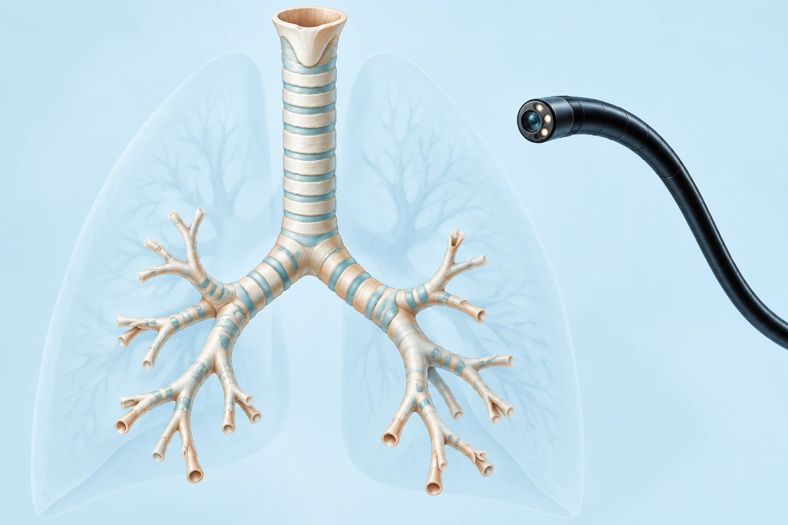
The windpipe is called the trachea, and the main airways branching into the lungs are called bronchi. Narrowing, masses or other changes can occur in these structures. A biopsy takes a tissue sample to clarify a diagnosis; being referred for a biopsy does not in itself mean cancer.
What does bronchoscopy show?
During bronchoscopy, a camera-equipped instrument is passed through the mouth or nose to examine the inside of the airways. Washing fluid or tissue samples may be collected when needed. Not every lung lesion can be reached by bronchoscopy; the method depends on the lesion’s location.
EBUS, mediastinoscopy and thoracoscopy
EBUS combines bronchoscopy with ultrasound to assess and sample lymph nodes next to the airways. Mediastinoscopy surgically accesses certain lymph nodes in the mediastinum. Thoracoscopy introduces a camera into the chest cavity to sample suitable tissues. These methods are not interchangeable.
Preparing for the procedure
Share previous scans, allergies and all medications with the team. In particular, do not stop blood thinners on your own; the procedural team determines fasting and medication instructions. If sedation or anesthesia is planned, discuss travel home and the need for someone to accompany you in advance.
Risks and interpretation of results
Depending on the procedure and sampling site, bleeding, infection, low oxygen levels or pneumothorax may occur. Pathology sometimes requires additional tests; an inadequate sample may lead to consideration of another method. Severe breathlessness or significant bleeding after the procedure requires emergency assessment.
Post-intubation tracheal stenosis
Scar tissue developing after intubation can narrow the windpipe. Increasing breathlessness and noisy breathing require assessment.
Tracheal stenosis: read about surgery and stent options →
Frequently asked questions
When will the biopsy result be ready?
This depends on the tests needed on the tissue; the laboratory examining the sample can provide the expected timing.
Does a negative result always rule out disease?
No. Sample adequacy and consistency with imaging findings are considered together.
Further reading
Patient information sources · Sources checked: 22 October 2026. Checking sources does not mean the text has been medically approved by the physician.